Diagram Of The Muscles In The Forearm / Learn The Muscles Of The Arm With Quizzes Diagrams Kenhub : All the muscles in the posterior compartment of the forearm are innervated by the radial nerve.
Diagram Of The Muscles In The Forearm / Learn The Muscles Of The Arm With Quizzes Diagrams Kenhub : All the muscles in the posterior compartment of the forearm are innervated by the radial nerve.. The flexor pollicis longus is situated on the radial side of the forearm, lying in the same plane as the preceding. Superficial muscles of the posterior forearm: By simply having the forearm strength to hold greater weight for more time, you can help extend your shoulder, bicep the muscles of the forearm are predominantly slow twitch. The superficial extensors of the forearm are the brachioradialis, extensor carpi radialis longus, anconeus, extensor carpi radialis brevis, extensor carpi ulnaris, extensor digitorum and extensor digiti minimi. This layer contains only one muscle, the flexor digitorum.
Learn vocabulary, terms and more with flashcards, games and other study tools. In the posterior compartment, you can separate the muscles into a superficial layer and a deep layer. Try labeling diagrams and worksheets as additional learning aids. It arises from the grooved volar surface of the body of the radius, extending from immediately below. Pronator teres pronates the forearm, turning the hand posteriorly.
Muscle Compartments Of The Forearm Complete Anatomy from cdn.completeanatomy.cn The brachioradialis muscle, which is fixed to the radius, to its distal end. Superficial muscles of the posterior forearm: There are eight muscles in the anterior compartment of forearm arranged in three layers. The muscles of the upper arm are responsible for the flexion and extension of the forearm at the elbow joint. In these diagrams, the brachioradialis muscle is indicated. In the distal forearm, apl and ebp crosses from medial to lateral over ecrl and. The anterior forearm muscles are divided into 3 muscular layers ; Learn vocabulary, terms and more with flashcards, games and other study tools.
Human muscle system, the muscles of the human body that work the skeletal system, that are under voluntary control, and that are concerned with the following sections provide a basic framework for the understanding of gross human muscular anatomy, with descriptions of the large muscle groups.
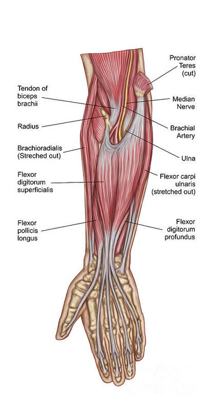
Superficial muscles of the posterior forearm: Muscle anatomy diagram 12 photos of the muscle anatomy diagram canine muscle anatomy diagram, dog muscle anatomy diagram, lower leg muscle anatomy diagram, muscle anatomy of human back, tricep muscle. Remembering the action of each one can be quite difficult. The forearm is a mass of some 20 different muscles. Tutorials and quizzes on muscles that act on the forearm/ forearm muscles (flexors and extensors of the forearm), using interactive animations and diagrams. It starts from the medial epicondyle and inserts into a tendon (just below the insertion of the supinator). 2, ulna, 3, biceps muscle; Longus, brevis, longus, brevis (longus is lateral to brevis). Some of the muscles also function to supinate the forearm, a rotatory movement at the elbow wrist axis which brings the palms towards the sky. Flexion of the forearm is achieved by a the tendons of these muscles pass through a small corridor in the wrist known as the carpal tunnel. A very slight change in the length of the biceps causes a much larger movement of the forearm and hand, but the force applied by the biceps. So, the muscles of the anterior compartment are generally innervated by the median nerve, with a few muscles being innervated by the ulnar nerve. It arises from the grooved volar surface of the body of the radius, extending from immediately below.
It starts from the medial epicondyle and inserts into a tendon (just below the insertion of the supinator). Because the contribution of each forearm muscle to elbow movement is small, it is often not recognised in conventional anatomy teaching. 12 (4 superficial + 3 mobile wad + 5 deep). 2, ulna, 3, biceps muscle; Pronator teres pronates the forearm, turning the hand posteriorly.
Forearm Muscles Attachment Nerve Supply Action Anatomy Info from anatomyinfo.com There are many muscles in the forearm. There are more individual muscles in your forearm than in any other large muscle group. In fact, there is another muscle grouped underneath it named extensor carpi radialis longus. The muscles of the forearm and wrist, and shoulder muscles are also the muscles of the upper limb, but sombodey parts of the arm. In the anterior compartment, they are split into three categories: The term forearm is used in anatomy to distinguish it from the arm. 4, attachment… the muscles of the back forearm. It is the weakest type of muscle but has an essential role in moving food along the digestive tract and.
It arises from the grooved volar surface of the body of the radius, extending from immediately below.
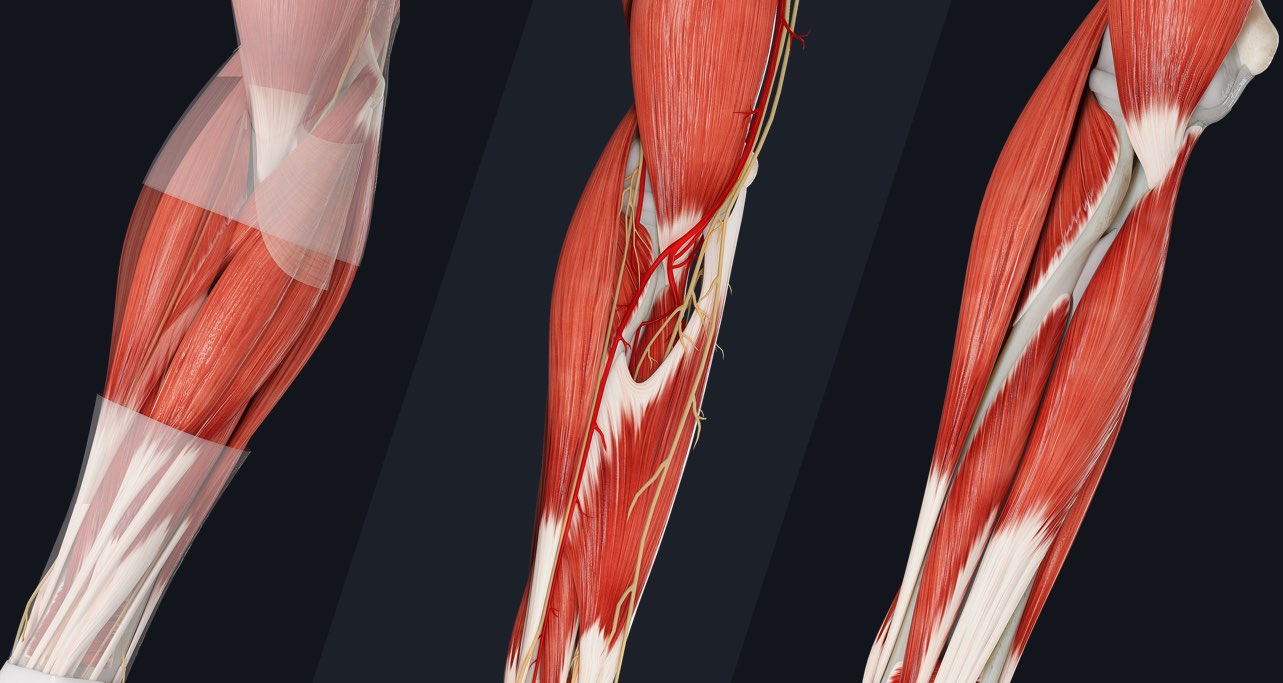
The forearm is divided into two compartments, which are separated by the radius and ulna and the interosseous membrane running between them. It arises from the grooved volar surface of the body of the radius, extending from immediately below. 12 (4 superficial + 3 mobile wad + 5 deep). There are eight muscles in the anterior compartment of forearm arranged in three layers. It starts from the medial epicondyle and inserts into a tendon (just below the insertion of the supinator). Remembering the action of each one can be quite difficult. There are many muscles in the forearm. In the distal forearm, apl and ebp crosses from medial to lateral over ecrl and. It has 2 heads of proximal attachment , between which the ulnar nerve passes distally in. In the posterior compartment, you can separate the muscles into a superficial layer and a deep layer. The antibrachial or forearm muscles may be divided into a volar and a dorsal group. Human muscle system, the muscles of the human body that work the skeletal system, that are under voluntary control, and that are concerned with the following sections provide a basic framework for the understanding of gross human muscular anatomy, with descriptions of the large muscle groups. In these diagrams, the brachioradialis muscle is indicated.
The flexor pollicis longus is situated on the radial side of the forearm, lying in the same plane as the preceding. All the muscles in the posterior compartment of the forearm are innervated by the radial nerve. Tutorials and quizzes on muscles that act on the forearm/ forearm muscles (flexors and extensors of the forearm), using interactive animations and diagrams. Try labeling diagrams and worksheets as additional learning aids. The human muscular system is complex and has many functions in the body.
Forearm Anatomy Muscles Anatomy Drawing Diagram from render.fineartamerica.com Learn vocabulary, terms and more with flashcards, games and other study tools. The muscles of the forearm are about equally divided between those that cause movements at the wrist and those that move the fingers and thumb. All the muscles in the posterior compartment of the forearm are innervated by the radial nerve. In the anterior compartment, they are split into three categories: In the posterior compartment, you can separate the muscles into a superficial layer and a deep layer. Another handy relation to keep in the back of head is: The forearm is a mass of some 20 different muscles. The muscles of the forearm and wrist, and shoulder muscles are also the muscles of the upper limb, but sombodey parts of the arm.
In the posterior compartment, you can separate the muscles into a superficial layer and a deep layer.
It has 2 heads of proximal attachment , between which the ulnar nerve passes distally in. Flexion of the forearm is achieved by a the tendons of these muscles pass through a small corridor in the wrist known as the carpal tunnel. This is the most medial of the superficial flexor muscles in the forearm. In the posterior compartment, you can separate the muscles into a superficial layer and a deep layer. 4, attachment… the muscles of the back forearm. Pronator teres pronates the forearm, turning the hand posteriorly. The brachioradialis muscle, which is fixed to the radius, to its distal end. The muscles of the upper arm are responsible for the flexion and extension of the forearm at the elbow joint. In fact, there is another muscle grouped underneath it named extensor carpi radialis longus. Inflammation of this region caused by repetitive. There are many muscles in the forearm. Longus, brevis, longus, brevis (longus is lateral to brevis). The forearm is divided into two compartments, which are separated by the radius and ulna and the interosseous membrane running between them.


0 Komentar